
Phase-based x-ray detection of threat objects – a new paradigm in security inspections?
By Professor Alessandro Olivo
X-rays are best known for use in security after healthcare (with personal belongings scanned at the airport before a flight). This use of x-ray is likely to increase in the near future: in some Chinese cities, for instance, bags are scanned at metro stations.
But the way x-ray images are created has remained practically the same for 120 years. Image formation relies on materials with different attenuation properties (the coefficient m) showing up differently subject to the amount of radiation passing through them (I1 and I2 in Fig. 1).
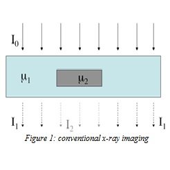
This approach is suboptimal when there are small differences in attenuation (because of similar characteristics in the “target” and its background or because the detail is very thin). This problem is well-known in medical imaging (since soft tissues have similar attenuation); but is encountered in all fields, including security inspections.
One way to overcome this limitation is to exploit the fact that x-rays are electromagnetic waves which change speed when moving through different materials. This leads to a wave front distortion, as parts of the wave front are “phase-shifted” with respect to others (figure 2(a)).
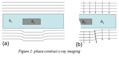
The physical quantity causing changes in x-ray speed (indicated with d, in figure 2) is much larger than that driving attenuation effects. If properly exploited, this can lead to much stronger image contrast. This is known as X-ray Phase Contrast Imaging.
But efforts to get a detector to see these changes in in speed have traditionally been extremely complex and extremely expensive, involving very specialized technology such as synchrotron radiation. There are only about 50 synchrotron facilities in the world; each costs >£100M, with the footprint of a football stadium. This does not suit the technique to everyday use in clinics or at airports!
At UCL, we have overcome this problem through observing that wave front distortions translate into changes in x-ray direction (see fig. 2(b)). This is effectively x-ray refraction just as, in visible light, a straw appears bent in a glass of water; but with x-rays, the deviations are measured in the micro-radian range: this is the equivalent to seeing a 1mm deflection from 1km away.
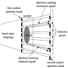
The good news is that systems to detect these miniscule shifts of angle are within reach. We place a pair of apertured masks either side of the imaged object with the two masks are slightly mismatched (see fig 3). This reduces the transmission of x-rays deviating in one direction, while increasing it for those going the other way. Small angular deviations are turned into significant intensity differences – even when a standard x-ray source is used.
![]()
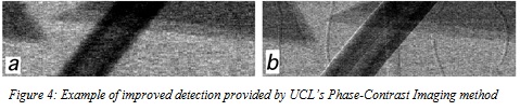
Through a project funded in 2007 by the Innovative Research Call (IRC) in Explosives and Weapons Detection (a cross-government programme sponsored by a number of Departments and Agencies under the UK Government’s CONTEST strategy), we showed this approach can lead to enhanced threat detection. Figure 4 shows a plastic cylinder, some thin wires and additional layers of material creating clutter: the wires are invisible in the conventional image (4a), but clearly detected in the Phase Contrast image (4b).
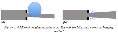
Recently, a more powerful effect has been observed, schematized in figure 5 (representing one of the “beamlets” created by the pre-sample mask from fig. 3). If the beamlet hits a detail that is larger than itself (5a), the entire beamlet is deviated; but if it strikes smaller details, multiple refraction occurs inside the beamlet, sending x-rays in different directions. These smaller events occur below the resolution limit of the imaging system, but a “global” effect (a broadening of the beamlet) can be observed and measured.
This is effectively a measure of the degree of inhomogeneity of the sample on the microscopic scale; for example, a perfectly homogenous sample would not produce any broadening. Preliminary evidence shows that this could help to distinguish between harmless and harmful materials (eg explosives), since it is unlikely for two materials to have the same microscopic structure even if they have similar attenuation characteristics. We could even assign a material-specific “tag” to shapes identified by these refraction signals (e.g. yellow-red for threat materials, blue-green for non-threat ones). This would easy for the operator to interpret and lend itself to automatic threat detection algorithms.
A new project funded by the IRC in Explosives and Weapons Detection 2013 initiative (a Cross-Government programme sponsored by a number of Departments and Agencies under the UK Government’s CONTEST strategy, in partnership with the US Department of Homeland Security, Science and Technology Directorate) is developing a demonstrator system, with the UCL team working in collaboration with Nikon Metrology. It is envisaged that, following a successful demonstration, prototype systems will be developed and subsequently commercialised and marketed by Nikon.
A new generation of more sensitive inspection systems could emerge from this, leading to improved security at airports and elsewhere.
Alessandro Olivo is professor of Applied Physics at the UCL Department of Medical Physics and Biomedical Engineering, and the Head of the X-Ray Phase Contrast Imaging Group. His research on phase-based x-ray imaging has been funded primarily by EPSRC (grants EP/I021884/1, EP/I022562/1, EP/L001381/1 and EP/G004250/1): this included funding (2009-13) under the Global Uncertainties Programme.
